New Back-Up Airway Debut - iGel How To
Straight to the Point:
Our department is about to embark on an exciting foray into cutting edge airway tools! The iGel will be deployed as the back-up airway of choice for our department starting on Monday, March 5. Please read on the familiarize yourselves with this absolutely vital device that will enhance our abilities to care for patients with difficult airways. This contains all necessary info regarding use of these new devices that we now wield in our airway expertise.
The Nitty Gritty:
What is it?
The iGel is a supraglottic airway that is made of malleable plastic that when heated to physiologic temperatures creates an anatomic seal over the hypopharynx and directs breaths into the larynx. It is NOT considered a definitive airway, but is a great device to use as a back-up should you have difficulty intubating on first-pass. It will replace our King Airways and LMA’s as the back-up airway of choice for our department.
Why are we switching?
The iGel is far simpler to use than most other back-up devices. It simply requires insertion and minimal management from there. There is no need to inflate balloon(s) (which in several animal and cadaveric studies have been shown to limit blood flow to the brain, less than ideal in many situations where back-up airways are needed). Further, you can pass an NG tube down the side port allowing you to decompress the stomach and further reducing the risk of aspiration. Lastly, it can be used to facilitate fiberoptic intubation (see below).
I cannot get this airway despite my best attempt(s)…quick, how do I use the iGel?
· Choose the correct size based on patient weight (we will carry size 3-5):
o Size 3 (yellow) = 30-60kg
o Size 4 (green) = 50-90kg
o Size 5 (orange) = 90kg+
· Place lubricant on the posterior surface (this is the part that will touch the palate)
· Simply scissor open the airway and slide it in the airway with the opening pointed toward the surface of the airway (aka insert with the opening towards the tongue)
· The device should seat fairly easily at which point hook up your EtCO2 monitor and BVM and bag away (your EtCO2 may be low but you should still a waveform if ventilating)
· There is a side port which can pass a 12Fr NG tube down for gastric decompression
· Breathe easy for a moment after what had to be a bit of a harrowing airway as you should be able to oxygenate and ventilate
· See attached video for a quick example of insertion by the skilled Dr. Longhurst!
Phew, that was easier than I thought. The iGel is in, what do I do from here?
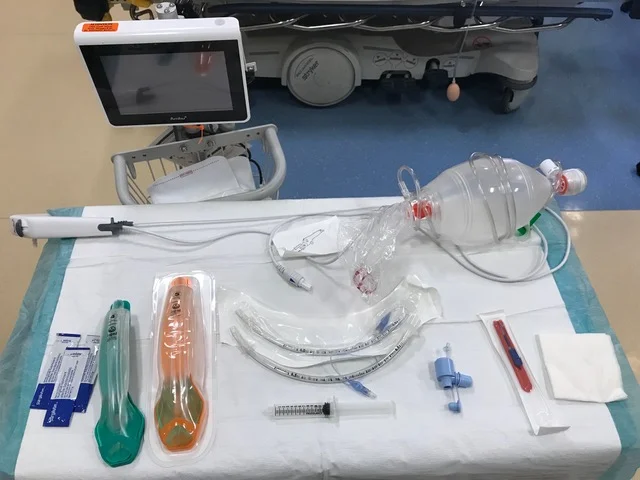
Assemble your supplies for fiberoptic intubation:
o More lubricant
o ETT – size it as 2.5 + LMA size (and be sure to have a size smaller)
o 10cc Syringe
o Bronchoscopy Adapter
o Fiberoptic Scope & Screen
o Cricothyrotomy supplies (at least a scalpel and gauze; it does not hurt to always be prepared)
o At least 2 sets of hands (ideally 3)
· Now, heavily lubricate your ETT and make sure the balloon is uninflated
· Advance the ETT to ~15cm into the LMA
· Place the bronchoscopy adapter onto the ETT and now you can oxygenate and ventilate throughout the process by bagging through the side port
· Advance the fiberoptic scope through the bronch adapter and tube. You should come out near the end of the LMA and visualize the cords right away. From there advance the fiberoptic scope through cords (essentially using it as a bougie)
· While the scope operator holds the scope in place, have an assistant advance your tube over the scope and inflate the balloon once the tube is visualized above the carina and remove the scope.
· At this point touchdown dances are in order as you officially have a definitive airway!
· If desired (and probably ideal since we are the airway experts in the hospital and many other services may not be familiar with an iGel), you can proceed to remove the iGel. To do this, remove the T-top of the ETT. Use the one size smaller ETT to stabilize the ETT in place and have an assistant gently back out the iGel. When possible grab the ETT proximally in the oropharynx as possible and hold it in place while the iGel is fully removed.
· Click here (https://www.dropbox.com/s/750e29cxmk5hm41/Fiberoptic%20Tube%20via%20iGel.MOV?dl=0) for a video for the entire procedure as demonstrated/narrated by Dr. Haas (who managed to master the technique after one practice run) with assistance by the strong but silent Dr. Vijitakula
Please take the time to familiarize yourself with these as they will save you in a pinch for any number of airway disasters. We must own this process as the emergent airway experts in the hospital. If you have any questions, do not hesitate to reach out.

