Intern Ultrasound of the Month: Acute Coronary Syndrome with Takotsubo Pattern
The Case
60s year old female with history of hypertension, hyperlipidemia, CVA presented to the emergency department for a few days of intermittent, non radiating substernal chest pressure with associated shortness of breath. Pain was more severe the day of presentation, thus prompting her to come to the ED. She denied any prior cardiac history and had never had pain like this before. Also denied any other current or recent symptoms.
On arrival, she was slightly hypertensive and tachycardic to 110s though not in any distress and appeared comfortable. Her exam was nonfocal.
Cardiac workup was initiated. EKG showed sinus tachycardia without conduction delay or ST elevation/depression but anterolateral T wave inversions were present. This was new compared to prior EKG.
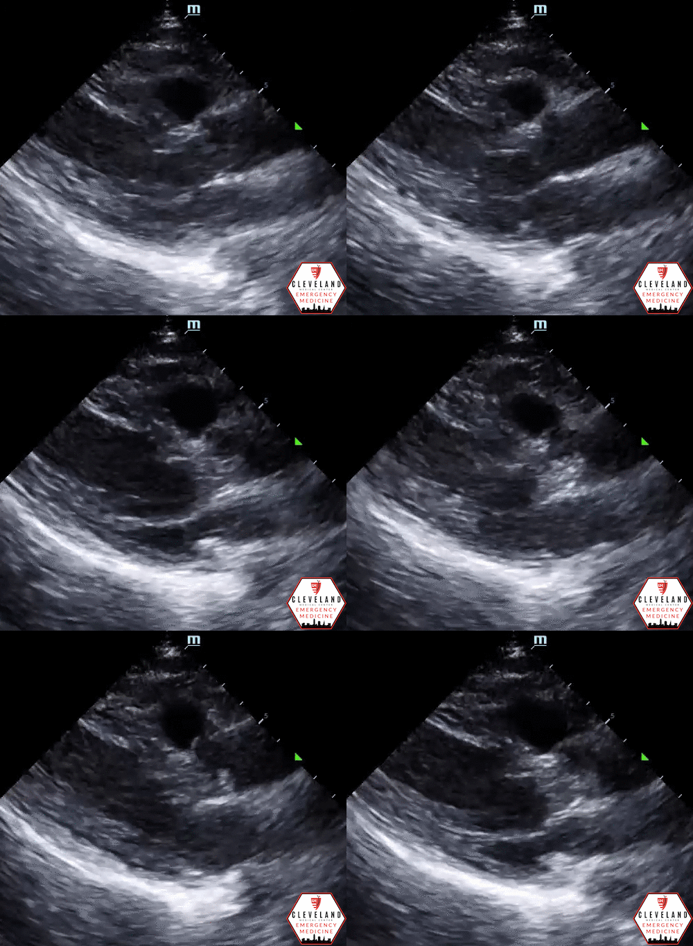
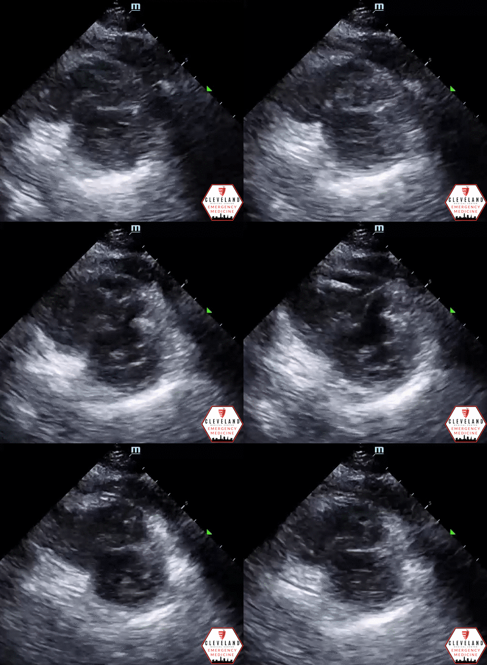
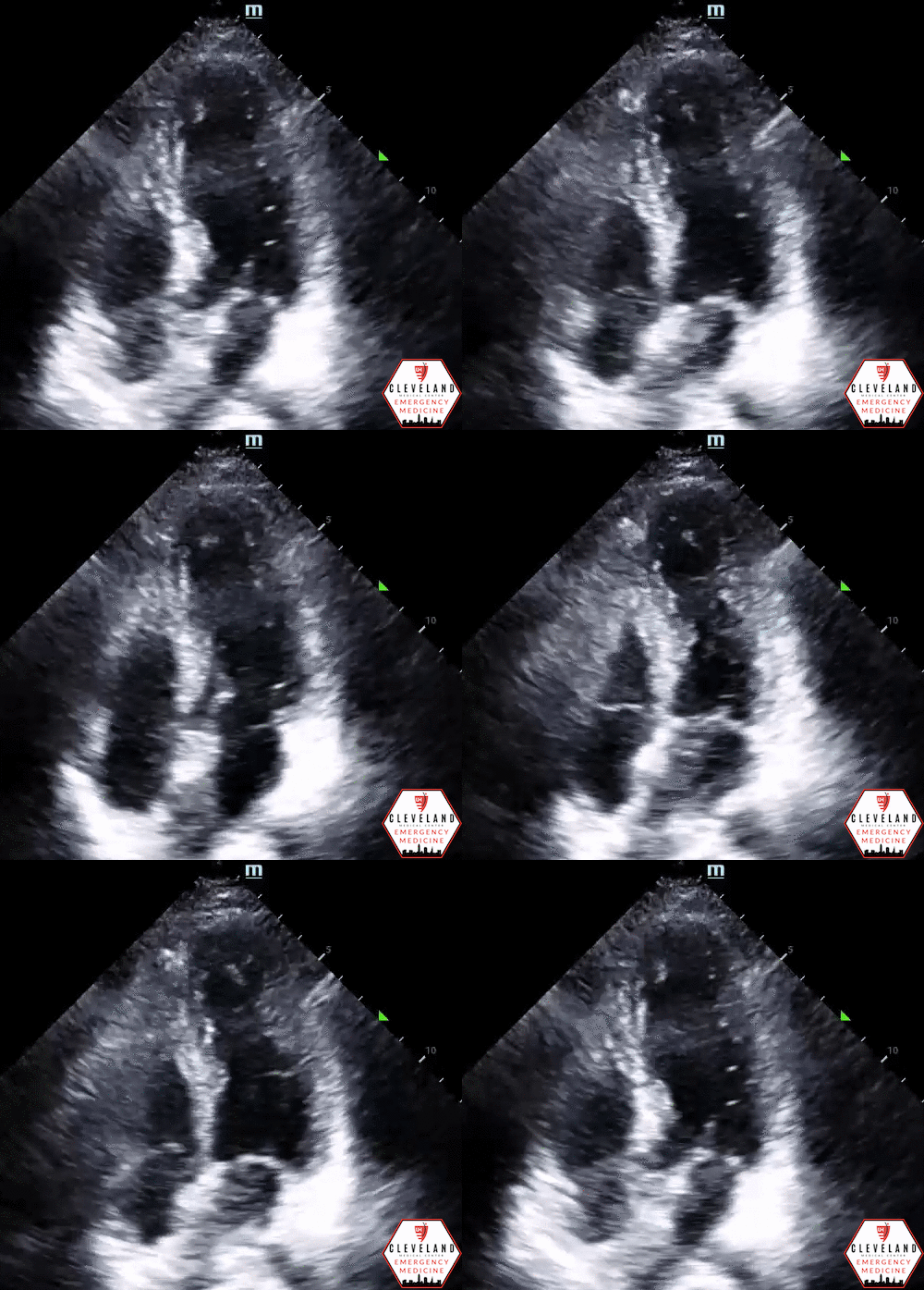
POCUS was performed…
POCUS findings: relatively preserved left ventricular basal function with apical ballooning and hypokinesis - a pattern consistent with classic Takotsubo but also highly concerning for acute coronary syndrome; new compared to prior echo. There is no pericardial effusion or signs of right heart strain.
Case continued: Labs were significant for troponin 1.5 as well as elevated VTE ddimer (CT PE was negative). Because of her symptoms and clinical findings (including new regional wall motion abnormalities on POCUS), cardiology was consulted from the ED. She received aspirin, plavix, and was started on a heparin drip. She was admitted to the cardiology service for cardiac cath the next day. In the meantime, her troponin peaked above 2, and comprehensive echo showed EF 35%, (normal two years ago). Left heart cath revealed multi-vessel disease with near occlusion of the RCA & LAD (the latter of which correlates with the apical hypokinesis). She underwent PCI with stents placed in both of these vessels. Ultimately did well overall and was discharged a few days later.
Cardiac POCUS & Assessment of Regional Wall Motion Abnormalities
Chest pain is a common presenting symptom in the emergency department. The vast majority of patients with acute coronary syndrome (ACS) have non-ST-segment elevation myocardial infarctions (NSTEMI) [1]. In the absence of diagnostic clinical findings, risk stratification and recognizing the need for prompt intervention can be difficult. POCUS assessment, particularly to evaluate for regional wall motion abnormalities (RWMA), has shown added diagnostic value for these patients and should be considered in any patient presenting with symptoms, history/risk factors, and clinical findings concerning for ACS.
Significance of RWMA
RWMA are among the earliest clinical manifestation of myocardial ischemia. Precedes EKG changes and onset of symptoms [2].
The degree of RWMA has been shown to correlate with degree of ischemia injury [3]
The presence of any RWMA is associated with increased risk of imminent adverse cardiac events [4].
When combined with history and clinical findings (including those with nondiagnostic EKGs), echo findings improve ability to diagnose ACS & can augment risk stratification [3, 5-6]
Emergency physicians have demonstrated the ability to detect RWMA in patients with coronary ischemia [3].
ACS vs Takotsubo
RWMA typically associated with ACS but can also be seen with Takotsubo cardiomyopathy
Takotsubo cardiomyopathy = transient stress-induced cardiomyopathy, commonly associated with emotional stress, that clinically mimics ACS but in the absence of coronary artery stenosis
Sonographically, this is classically seen as apical LV ballooning with akinesis/hypokinesis, although variant forms exist
Differentiation between the two is difficult & requires cardiac catheterization to definitively diagnose [7-9].
POCUS Assessment of RWMA
3-segment assessment & its anatomical correlates — a simplified version of the 17 segment quantitative assessment proposed by the American Heart Association [10-11]. Focused echo by emergency physicians using this assessment has demonstrated good utility in detecting RWMA [3]
LV segment correlation with coronary artery perfusion
Anterior wall = Left anterior descending artery (LAD)
Inferior wall = Right coronary artery (RCA)
Lateral wall = Left circumflex (L Cx)
Coronary artery distributions on EKG
Coronary artery distributions seen on cardiac ultrasound
What is considered a RWMA?
Relative hypokinesis, dyskinesis, akinesis of an LV segment compared to the rest of the LV
May appear as asymmetric contractility or myocardial thinning
How to evaluate for RWMA?
Obtain multiple cardiac views, visualizing the entire LV, for a thorough, more reliable assessment. Qualitatively assess for abnormal LV wall segment contractility/thinning.
The parasternal short axis view at the level of the papillary muscles (see image above) is thought to be the best view as it allows for simultaneous visualization of the different walls.
*If not at this level or slightly off-axis, the assessment may appear falsely normal or abnormal.
**For this patient, however, the apical view was most significant
A few pitfalls:
Suboptimal images -- body habitus, anatomy, positioning
Off-axis views can lead to false positive or negative assessments
Pre-existing or concurrent pathology may confound the assessment —> helpful to compare to a previous echo [12].
Take Home Points
In combination with patient history & clinical findings (i.e. EKG, troponin, etc) , cardiac POCUS adds significant diagnostic value in evaluating patients for ACS & helps with risk stratification.
RWMA may appear as asymmetric contractility and/or myocardial thinning. POCUS findings may vary depending on which vessel(s) involved. Correlate findings with EKG changes & prior echos to help determine acuity
PSSA at the level of the papillary muscles is thought to be the best view to assess for RWMA. But, as always, it’s important to obtain multiple views (as demonstrated in this case).
Be familiar with Takotsubo findings but treat as ACS until proven otherwise with a clean cath
POCUS findings can expedite cardiology involvement and further workup (such as comprehensive echo, catheterization, etc.)
POST BY: DR. JEHANNE BELANGE, PGY1
FACULTY EDITING BY: DR. LAUREN MCCAFFERTY
References
Amsterdam E, Wenger N, Brindis R, et al. 2014 AHA/ACC guideline for the management of patients with non-ST-elevtion acute coronary syndromes: A report of the american college of cardiology/american heart association task force on practice guidelines. Circulation. 2014;130:344-426.
Hauser A, Vellappillil G, Ramos R, Gordon S, Timmis G, Dudlets P. Sequence of mechanical, electrocardiographic and clinical effects of repeated coronary artery occlusion in human beings: Echocardiographic observations during coronary angioplasty. J Am Coll Cardiol.. 1985;5:193-197.
Frenkel O, Riguzzi C, Nagdev A. Identification of high-risk patients with acute coronary syndrome using point-of-care echocardiography in the ED. Am J Emerg Med. 2014;32:670-672.
Sabia P, Afrookteh A, Touchstone D, Keller M, Esquivel L, Kaul S. Value of regional Wall Motion Abnormality in the emergency room diagnosis of acute myocardial infarction A Prospective study using two-dimensional echocardiography Circulation. 1991;84:85-92.
Kontos MC, Arrowood JA, Paulsen WH, et al. Early echocardiography can predict cardiac events in emergency department patients with chest pain. Ann Emerg Med. 1998;31:550-557.
Ha, E. T., Cohen, M., Fields, P. J., Daele, J. V., & Gaeta, T. J. (2019). The Utility of Echocardiography for Non-ST-Segment Elevation Myocardial Infarction: A Retrospective Study. Journal of Diagnostic Medical Sonography, 36(2), 121-129
Bybee KA, Kara T, Prasad A, et al. Systematic Review: Transient Left Ventricular Apical Ballooning: A Syndrome That Mimics ST-Segment Elevation Myocardial Infarction. Annals. 2004; 141(11): 858-865.
Gianni M, Dentali F, Grandi AM, Sumner G, Hiralal R, Lonn E. Apical ballooning syndrome or takotsubo cardiomyopathy: a systematic review. Eur Heart J. 2006 Jul;27(13):1523-9.
Nguyen TH, Horowitz, JD. Differentiating Takotsubo cardiomyopathy from myocardial infarction. eJ Cardiol Practice. 2014; 13(7). Retrieved from https://www.escardio.org/Journals/E-Journal-of-Cardiology-Practice/Volume-13/Differentiating-Tako-tsubo-cardiomyopathy-from-myocardial-infarction
Johnson B, Lovallo E, Frenkel O, Nagdev A. Detect cardiac regional wall motion abnormalities by point-of-care echocardiography. https://www.acepnow.com/article/detect-cardiac-regional-wall-motion-abnormalities-point-care-echocardiography/. Accessed 1/26/2021..
Cerqueira M, Weissman N, Dilsizian V, et al. Standardized myocardial segmentation and nomenclature for tomographic imaging of the heart A statement for healthcare professionals from the cardiac imaging committee of the council on clinical cardiology of the american heart association Circulation. 2002;539-542.
Ma OJ, Mateer JR, Reardon RF, & Joing S. (2014). Ma and Mateer's Emergency Ultrasound. New York, NY: McGraw-Hill Education.


