Intern Ultrasound of the Month: Utility of POCUS for 1st Trimester Bleeding in the ED
The Case
29-year-old female, G6P3023 at approximately 7 weeks gestation by LMP, presented to the emergency department for 3 days of vaginal bleeding and abdominal cramping. She stated she was in her normal state of health when she became nauseated then started having intermittent episodes of spotting and vaginal bleeding. She denied the passage of clots or fetal parts or any vaginal discharge associated with the bleeding. She also reported abdominal cramping in the upper quadrants bilaterally but no lower abdominal pain. She reported two prior miscarriages but noted that this felt different than those pregnancies. Her review of systems was otherwise negative and she had no other comorbidities or prior surgeries. She had not yet seen OB during this pregnancy.
Her vitals were within normal limits and abdominal exam was unremarkable. Pelvic exam showed no active bleeding, cervix was closed. The rest of the physical exam was noncontributory.
Point-of-care ultrasound was performed to evaluate for intrauterine pregnancy (IUP).
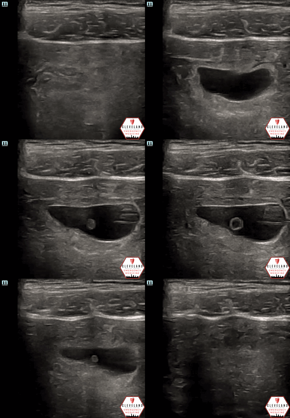
Curvilinear probe
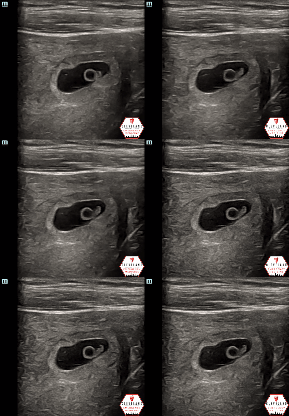
Linear probe
POCUS Findings:
An intrauterine gestational sac was visualized with the transabdominal probe without obvious contents within it. However, with a high-frequency linear probe, a yolk sac and fetal pole was visualized, which confirmed an IUP. Flickering within the fetal pole suggested fetal heart beat. No free fluid was seen.
Case Continued:
Intrauterine pregnancy (IUP) was confirmed, thus significantly reducing the likelihood of an ectopic pregnancy. She was Rh+ so RhoGam was not indicated, and CBC showed a normal hemoglobin level. She was counseled on threatened spontaneous abortions and discharged home with plans for close OB follow up.
POCUS in the Assessment & Management of First Trimester Vaginal Bleeding in the ED
Table 1. Classification of early pregnancy loss [2].
Vaginal bleeding occurs in 20-40% of first trimester pregnancies [1]. As emergency physicians, it is essential that we appropriately and efficiently evaluate patients presenting with bleeding and accurately recognize (potential) signs of pregnancy loss - see Table 1 [2]. Point-of-care ultrasound (POCUS) has become an invaluable diagnostic tool for the workup and management of these patients in the emergency department (ED) [3-6].
The primary objective of POCUS in the first trimester is to confirm an intrauterine pregnancy, which essentially excludes ectopic pregnancy (unless there’s higher risk for heterotopic pregnancy, such as with in-vitro fertilization). Secondary objectives are to assess viability of an intrauterine pregnancy and evaluate for free fluid. Additionally, other signs of ectopic pregnancy (i.e. adnexal mass, extrauterine gestational sac +/- yolk sac or fetal pole) may be detected; however, these are not always visualized and their absence does not rule out an ectopic [7-9].
Transabdominal POCUS is widely available and should be the first line imaging modality for pregnant patients presenting with vaginal bleeding. If an IUP is confirmed, patients can generally be discharged with outpatient OB follow-up. If an IUP is not confirmed with a transabdominal POCUS, this should be considered an ectopic pregnancy until proven otherwise, even though it may still be an early intrauterine pregnancy. At this point, a transvaginal ultrasound (POCUS vs radiology-performed) should be performed as it allows for higher resolution and better visualization of structures, though is more invasive [5].
Multiple studies have shown that emergency physicians can accurately and rapidly evaluate for an intrauterine pregnancy with POCUS with specificity approaching 100%. POCUS also reduces the rate of missed ectopic pregnancies, expedites definitive management for ectopic pregnancy, and decreases length of stay [3-4].
What about b-HCG levels & the “discriminatory zone”?
The discriminatory zone is the b-HCG level above which an IUP is expected to be seen on ultrasound. While HCG levels may correlate with IUP determination for normal intrauterine pregnancies, this is not always reliable, particularly for extrauterine pregnancies [3-4]. In a cross sectional study by Wang et al, the previously reported discriminatory zone had very poor sensitivity and specificity, and it wasn’t until the HCG level exceeded 25,000 that an IUP was identified in the vast majority of patients [4]. If there is any concern for ectopic pregnancy (i.e. IUP has not yet been confirmed), imaging or management decisions should NOT be determined by HCG level alone.
Review of the POCUS Assessment for First Trimester Vaginal Bleeding
To perform a transabdominal OB scan, place a 3.5 mHz curvilinear probe just above the pubic symphysis. With the probe indicator pointing toward the patient’s head, evaluate the pelvis in the sagittal plane. Locate the endometrial stripe, an echogenic line in the center of the uterus. Fan the probe from side of the pelvis to the other, visualizing the entirety of the uterus, bladder, and surrounding adnexa to evaluate for signs of intrauterine pregnancy, free fluid, or other major abnormalities. Make sure you have adequate depth, i.e. you can visualize at least a few centimeters beyond the deepest aspect of the uterus, to ensure an adequate assessment. Then, to obtain a transverse view, rotate the probe 90 degrees counterclockwise so the indicator is pointing toward the patient’s right. Again identify the endometrial stripe in the center of the uterus and fan the probe through the entire uterus from fundus to cervix [8-9].
What confirms an IUP?
A yolk sac or fetal pole within the intrauterine gestational sac must be present in order to confirm an IUP. A gestational sac alone is not enough, as a pseudogestational sac may accompany an ectopic pregnancy. Additionally, gestational sac should lie within the endometrial cavity; if more eccentrically located, measure the endomyometrial mantle — if less than 5-8 cm, this could indicate an interstitial or cornual ectopic pregnancy (despite being within the uterus) [7-9]. See Table 2 for general trend of development in early pregnancy.
Table 2. General trend of development in early pregnancy [8]
Once an IUP is visualized, what’s next?
Determine viability by assessing fetal cardiac activity. Using m-mode, place the bar over the fetal heart beat, generate a tracing, and measure one cycle to the next (your machine should calculate the heart rate). If you can see cardiac activity but not getting a clear tracing, try slowing down the speed (method varies by machine) as this may yield a better waveform. If unable to visualize a fetal heart beat, it may too early or it may indicate fetal demise. While m-mode (and color doppler) are safe to the fetus, avoid use of pulse wave doppler as this transmits more acoustic energy to the fetus which could be harmful [8-9].
Once an IUP is confirmed, no additional imaging or HCG level is generally indicated unless other complicating factors are present.
What if no IUP is visualized?
If no IUP is visualized, this is an ectopic pregnancy until proven otherwise, and a FAST exam should be performed. In the setting of suspected ectopic, the presence of free fluid, particularly in the right upper quadrant, predicts the need for operative intervention [6]. Other signs of ectopic pregnancy, such as extrauterine yolk sac/fetal pole or adnexal mass, may be seen but are often not [7-9].
The Utility of a High-Frequency Linear Transducer
In pregnant patients with ~5-8 weeks gestational age, it may be difficult to clearly visualize an IUP with the commonly used low frequency probe. Based on a study by Tabbut et al, a high-frequency linear probe allowed for IUP confirmation in 33% of patients whose scans with a curvilinear probe were indeterminate. Additionally, of the 66% of pregnancies that could not be confirmed, 83% could also not be visualized by a transvaginal ultrasound exam and, thus, were most likely ectopic pregnancies [10]. While this was only a pilot study, these results suggest that a high-frequency linear probe may be useful in patients with first trimester vaginal bleeding and may significantly reduce the amount of transvaginal scans required in the ED. This reduction may ultimately help save the patient and the ED time, cost, and potential stress/complications from the procedure itself. A downside of the linear probe is that it is limited by body habitus due to depth restrictions.
Similar to the case above, these images illustrate the improved image quality with the linear probe (right) compared to the curvilinear probe (left)
Curvilinear transducer not able to detect IUP [5]
Linear transducer able to detect pregnancy at 7 weeks [5]
A Few Other Pearls and Pitfalls:
Transabdominal OB scans are significantly improved if the patient has a full bladder as this provides an acoustic window for better visualization of deeper structures. Transvaginal ultrasound, on the other hand, is better with an empty bladder.
If you can’t quite confirm IUP due to image quality, consider decreasing your depth to allow for better visualization of the structures of interest; this tends to produce better quality images than simply zooming in.
Differentiate free fluid from anatomic structures based on shape. Free fluid has sharper and irregular edges whereas cysts and vessels have a more well-defined shape.
Don’t forget the basics! Even if you immediately see an IUP or abnormal structures, remember to evaluate the pelvis in both sagittal and transverse planes and fan through all the structures completely. You never know what else you might find!
Take Home Points:
Always attempt transabdominal POCUS to evaluate for IUP and free fluid. Findings can expedite your management and disposition.
A yolk sac or fetal pole within the gestational sac in the uterus must be present to confirm an IUP.
Lack of IUP with a positive pregnancy test is an ectopic pregnancy until proven otherwise. Consider a FAST exam; the presence of free fluid should raise your concern even further, especially if present in the RUQ.
If unable to confirm IUP with the low frequency probe, consider using a high-frequency linear transducer as this may reduce the need for transvaginal ultrasound (which can save the patient time, money, and discomfort).
POST BY: AUSTIN SCHOEFFLER, MD (R1)
FACULTY CO-AUTHOR/EDITOR: LAUREN MCCAFFERTY, MD
References
Ankum WM, Van der Veen F, Hamerlynck JV, Lammes FB. Suspected ectopic pregnancy. What to do when human chorionic gonadotropin levels are below the discriminatory zone. J Reprod Med. 1995;40:525–8.
Vaginal bleeding in pregnancy (less than 20wks). WikEM. (n.d.). Retrieved September 20, 2022, from https://wikem.org/wiki/Vaginal_bleeding_in_pregnancy_(less_than_20wks)
McRae A, Murray H, Edmonds M. Diagnostic accuracy and clinical utility of emergency department targeted ultrasonography in the evaluation of first-trimester pelvic pain and bleeding: a systematic review. CJEM. 2009;11(4):355-64.
Wang R, Reynolds TA, West HH, Ravikumar D, Martinez C, McAlpline I, et al. Use of a β-hCG discriminatory zone with bedside pelvic ultrasonography. Ann Emerg Med. 2011; 58(1)12-20.
Panebianco NL, Shofer F, Fields M, Anderson K, Mangili A, Matsuura AC, et al. The utility of transvaginal ultrasound in the ED evaluation of complications of first trimester pregnancy. Am J Emerg Med. 2015; 33: 743–748
Moore C, Todd WM, O’Brien E, Lin H. Free fluid in Morison’s pouch on bedside ultrasound predicts need for operative intervention in suspected ectopic pregnancy. Acad Emerg Med. 2007;14(8):755-8.
Dighe M, Cuevas C., Moshiri M, Dubinsky T, Dogra VS. Sonography in first trimester bleeding. J Clin Ultrasound. 2008; 36(6), 352–366.
Reardon RF, Hess-Keenan J, Roline CE, Caroon LV, Joing SA. (2014). First Trimester Pregnancy. In OJ Ma, JR Mateer, RF Reardon, SA Joing (eds), Ma and Mateer’s Emergency Ultrasound (3rd ed). New York, NY: McGraw-Hill Education. pp 381-424.
Noble V, Nelson B. Manual of Emergency Medicine and Critical Care Ultrasound, 2nd ed. Cambridge: Cambridge UP, 2011.
Tabbut M., Harper D., Gramer D., Jones R. High-frequency linear transducer improves detection of an intrauterine pregnancy in first-trimester ultrasonography. Am J Emergency Med. 2016; 34(2), 288–291.


