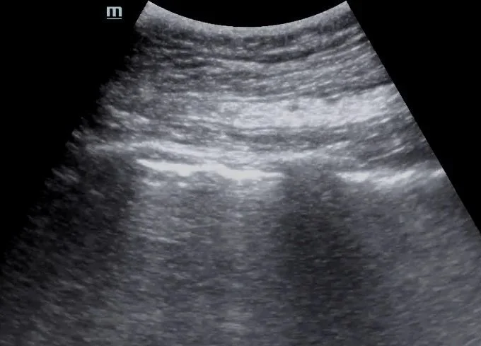
This post by Dr. Jordan DeAngelis reviews the use of point-of-care lung ultrasound to assess for pneumothorax, focusing on image acquisition, interpretation of lung sliding and the lung point, and practical tips to avoid common pitfalls.
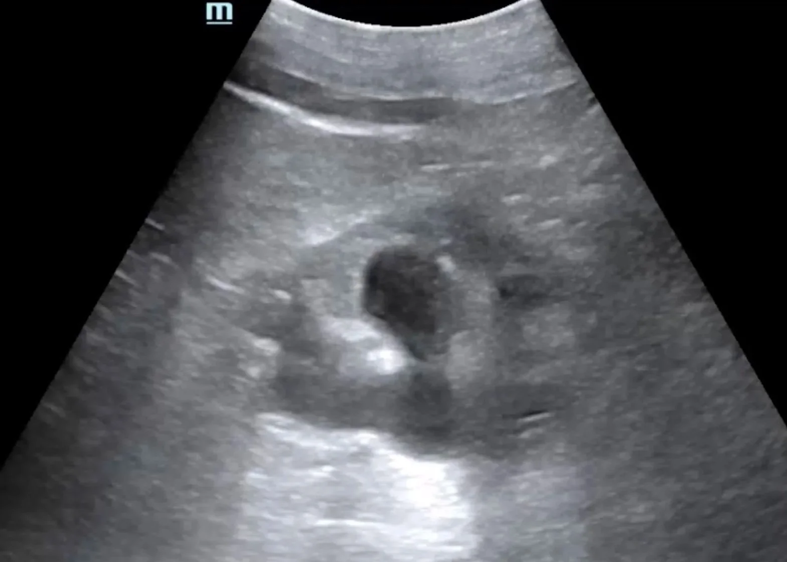
This case is by Dr. Nicole Minnerath and illustrates the detection of new hydronephrosis in a patient with bilateral nephrostomy tubes and ureteral stents. Read on to explore the role of renal and bladder POCUS in evaluating obstructive uropathy.
In this post, Dr. Tiana Sarsour highlights a case of recurrent abscesses and fever where POCUS confirmed a drainable collection, followed by a great review of abscess management and the role of ultrasound for skin and soft tissue infections.
This case is by Dr. Hannah Hendrix (now PGY2) and features a great example of the retrobulbar spot sign — a unique finding associated with embolic central retinal artery occlusion (CRAO). Read on for a quick review of ocular ultrasound and a closer look at this highly specific sonographic feature.
This post by Dr. Thomas Evans walks through the essentials of ultrasound-guided pericardiocentesis — when to do it, how to do it, and what pitfalls to avoid. We highlight the subxiphoid, parasternal, and apical approaches with practical POCUS tips for real-time needle visualization, plus a review of complications and how to keep the procedure as safe as possible.
This case is by Dr. Ryan Starkman (now PGY2) and is a great example of a large circumferential pericardial effusion with sonographic tamponade physiology diagnosed with POCUS. This expedited interventional cardiology involvement before the patient decompensated, and 750cc of fluid was successfully drained in the cath lab.
This case is by Dr. Jacob Perino and is a great example of an incidental finding of an iliac aneurysm found when performing a bladder ultrasound on a patient presenting with a malfunctioning Foley catheter. Read on to learn more about iliac aneurysms and bladder ultrasound in the setting of a Foley.
This month’s case is by Dr. Yanina Guevara who shares a case of painless monocular vision changes in a patient who was found to have a retinal detachment (and vitreous hemorrhage), first diagnosed with point-of-care ultrasound! This is accompanied by an overview of how to perform an ultrasound ocular exam and highlights some of the pathologies that can be visualized. Read on to learn more!
Our latest Intern Ultrasound of the Month is by Dr. Abby Wissman and features a cool case of a complex cystic renal mass found incidentally. She reviews the basics of renal POCUS and dives into a great discussion about various renal pathology that we may see on ultrasound.
To follow up our most recent case of McConnell’s sign in the setting of an acute pulmonary embolism, Dr. Isma Dhanani goes more in-depth on the right heart and covers additional signs of right heart dysfunction - acute and chronic - that can be quickly identified with POCUS.
Our latest Intern Ultrasound of the Month is brought to you by Dr. Gabe Alagna. He presents a case in which POCUS performed on a patient in cardiac arrest found an enlarged right heart with McConnell’s sign, raising concern for PE, which quickly changed management. Read on to learn more!
Our latest Intern Ultrasound of the Month is by Dr. Annie Thai. She shares a case of a symptomatic abdominal aortic aneurysm and provides a great review of how to perform and optimize your aorta ultrasound exam. We hope you enjoy!
Our next Intern Ultrasound of the Month is by Dr. Dave Wiliams and features a classic case of cellulitis in which POCUS helped support the clinical diagnosis and rule out an abscess. Read on for a great review of cellulitis and how to evaluate for soft tissue infections using POCUS.
Our next Intern Ultrasound of the Month is by Dr. Matthew Hom and features a great case of vitreous hemorrhage diagnosed with POCUS. Read on to learn more about this and how to differentiate from ophthalmologic emergencies such as retinal detachment.
Our next Intern Ultrasound of the Month is by (now PGY2) Dr. Brian Fort and features a great case of post-viral pneumonia with associated pleural effusions diagnosed with POCUS when the patient was too unstable for CT. The patient’s course was further complicated by bacterial superinfection resulting in Lemierre syndrome.
Our latest Intern Ultrasound of the Month by Dr. Shruti Africawala features a great case of left ventricular hypertrophy and hypertrophic cardiomyopathy, along with a review of evaluating LV function with POCUS.
Not all hydronephrosis is a kidney stone! Our latest Intern Ultrasound of the Month by Dr. Brooke Ott features a great case of a patient presenting with flank pain whose renal POCUS found hydronephrosis in addition to complex mass in her pelvis. This led to the discovery of tubo-ovarian abscess as the culprit. Learn more in this blog post!
Flashes & floaters & POCUS, oh my! Our latest Intern Ultrasound of the Month is by Dr. Ranjana (RJ) Ravikumar and features a great case of a large retinal detachment diagnosed with POCUS! Check it out !
Our next Intern Ultrasound of the Month is by Dr. Kelsie Rhyne and features a great case discussion about the diagnostic and procedural utility of POCUS for evaluating and managing a painful, swollen knee (and other joints).
Our latest Intern Ultrasound of the Month by Dr. Vincent Marshall features a case of achilles tendon tear confirmed with POCUS. Read on to learn more!
Our latest Intern Ultrasound of the Month by Dr. Polly Wiltz features a case of ventricular tachycardia, visualized on POCUS, followed by a discussion on using POCUS in cardiac arrest. Read on to learn more!
Don’t just get a chest x-ray! Use lung ultrasound to improve your diagnostic ability! Our latest Intern Ultrasound of the Month by Dr. Ian Brallier features an interesting case of a complex pleural effusion with extensive lung consolidation from lung cancer. Read on to learn more about evaluating for pleural effusions (and other pathology) using ultrasound!
Did you know you can diagnose ligament injuries of the knee using POCUS? Our latest Intern Ultrasound of the Month by Dr. Shauna Combs features a case of an MCL tear and traumatic effusion followed by discussion on how to evaluate for various musculoskeletal injuries of the knee! Read on to learn more!
Our next Intern Ultrasound of the Month is a great review of POCUS for First Trimester Pregnancy by Dr. Austin Schoeffler!
Our first Intern Ultrasound of the Month for this academic year is by Dr. Auryana DeChick! It features a case of an incidentally found left atrial aneurysm as well as a great primer on acquiring the different cardiac views for a focused echo!
This latest Intern US of the Month is by Dr. Sam Hertz and features a great discussion on using POCUS to evaluate for ascites and guide paracentesis.
This latest Intern US of the Month case by Dr. Juan Valdes Infante showcases the utility of POCUS in evaluating for multiple ankle pathologies. Learn more about calcaneal avulsion fractures, which are often a surgical emergency, and how to assess for various ankle pathologies using POCUS!
This Intern US of the Month features a great case by Dr. Rich Dowd of an extensive aortic dissection spanning from the aortic arch to the bifurcation with extension into the left subclavian artery. Check out the full post to learn more!
This latest Intern US of the Month features a case of a traumatic pneumothorax and a great discussion on this time-saving and highly useful POCUS assessment by Dr. Blake Nelson!
This Intern US of the Month features a unique case of asteroid hyalosis, a mimicker of vitreous hemorrhage, diagnosed with POCUS by Dr. Eniola Gros!


