Intern Ultrasound of the Month: A Rare Complex Cystic Renal Mass
The Case
90-year-old male with past medical history including dementia presented to the ED after multiple unwitnessed falls at his skilled nursing facility. He was having difficulty ambulating due to pain in his left hip and thigh. He was only oriented to self so additional history was limited. His exam did not show obvious deformity or ecchymosis but he was tender to palpation along his left hip. His vitals were stable and he was neurovascularly intact without any other notable exam findings.
Plain films of his chest, hip, and femur were obtained, along with CT head and C-spine, all of which were negative for acute pathology. A CT abdomen/pelvis was also ordered to evaluate for occult hip fracture as well as intra-abdominal pathology given his falls and limited history. Labs were unremarkable aside from his urinalysis which was suggestive of a urinary tract infection and a large amount of blood. He did have frank hematuria as well.
While awaiting CT, point-of-care ultrasound of the kidneys and FAST exam was performed…
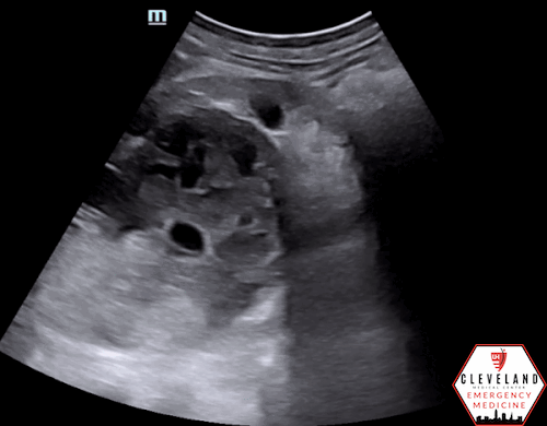
Sagittal view of the right upper quadrant with a large complex mass seen extending from the superior pole of the kidney

Transverse view of the kidney and mass

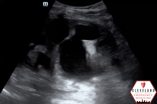
Color doppler over the mass showing no apparent internal flow
POCUS findings: In the right upper quadrant, a large complex cystic structure was seen extending from the superior pole of the right kidney. It has mixed echogenicity with hypoechoic areas suggestive of fluid. It appears contained and no free fluid is seen. Color doppler shows no evidence of internal flow. His other kidney was normal-appearing and his bladder was decompressed.

Case Continued: CT was read as a large (9 x 9 x 8 cm) complex, mostly cystic mass in the right kidney with thickened walls with nodularity and internal debris with infiltration into the perinephric fat along with fluid. This was concerning for a complex mass versus hematoma in the setting of trauma. Ultimately, radiology and urology felt that this was most likely a rare cystic renal cell carcinoma. He was admitted for pain control, physical and occupational therapy, and facilitation of further workup.
Renal POCUS
Image Acquisition (1)
Right kidney: The right kidney may be found by placing the probe in the mid-axillary line with the indicator pointing toward the patient's head, then sliding inferior to the liver until a characteristic “bean” shape is found. The kidney is typically seen around the 10th-11th intercostal space. You may need to rotate your probe to slide in between rib spaces and better visualize the kidney. In this view, you should be able to visualize the renal cortex, parenchyma, renal pelvis, renal sinus, major and minor calyces. Once you have an adequate view of structures, fan anteriorly and posteriorly through the entire kidney to help ensure you do not miss any major abnormalities. While maintaining this view, you may then rotate your probe 90 degrees to obtain a transverse view and fan through once again. In addition, color doppler should be applied to help differentiate prominent vasculature from hydronephrosis or other abnormalities such as cysts.
Left kidney: The major difference when moving to the left kidney is the initial placement of the probe, as the left kidney is more superior and posterior compared to the right kidney. The probe should be placed around the level of ribs 8-10 and in the posterior axillary line, which often requires your knuckles to touch the bed. Fan through the entire kidney in a longitudinal orientation then rotate the probe 90 degrees for a transverse view. Also remember to apply Color Doppler.
Bladder: It is important to evaluate the bladder as part of a renal ultrasound to assess for urinary retention and potentially identify a cause for renal pathology.

Figure 1. Renal anatomy (longitudinal orientation) with corresponding ultrasound image. Source: Koratala et al (2)

Figure 2. Importance of Color Doppler — A central hypoechoic area is concerning for possible hydronephrosis but Color Doppler indicates prominent renal vasculature. Source: NephroPOCUS (3)
Hydronephrosis
Evaluation for hydronephrosis is the most common application for renal POCUS. Hydronephrosis is an abnormal dilation of the kidney resulting from obstructive uropathy, caused by kidney/ureteral/bladder stones, masses, prostate enlargement, dysfunctional bladder, among others. It appears as a hypoechoic area in the kidney starting centrally and extending peripherally with greater degrees of severity (2), see Figure 3. As mentioned above, applying Color Doppler can help differentiate hydronephrosis from prominent renal vasculature (1). While less sensitive than CT, POCUS is a great initial diagnostic test with comparable diagnostic accuracy to radiology-performed ultrasound and shorter length-of-stay (4).

Figure 3. Hydronephrosis Grading. Source: POCUS 101 (1)

Figure 4. Progression of hydronephrosis sonographically. 4a. Mild hydronephrosis

4b. Moderate hydronephrosis
4c. Severe hydronephrosis
While evaluating for the presence of hydronephrosis is the primary focus of POCUS studies, it is important to recognize other pathology that may be easily visualized. When you see abnormal findings, take a closer look and correlate with the clinical context.

Figure 5. Simple cysts
Renal Cysts
Renal cysts are a relatively common and often benign pathology among hypoechoic kidney lesions. It is estimated that about 50% of people over the age of 50 have some form of cystic renal disease (5). A simple cyst is defined by ultrasound as a distinct circular mass of homogeneous anechoic content with smooth, well-defined margins with a barely detectable submillimeter wall, see Figure 5. In addition, the interface between the cyst and surrounding structures must be simple without a wall-off appearance. If these criteria are not met, then the lesion cannot be considered simple and instead moves into the atypical or complex category. Complex cysts often have internal septations, thickened irregular walls, or nodularity. Ultrasound can detect these findings with high sensitivity, sometimes outperforming CT (6). However, these atypical lesions are often incidentally identified and characterized by CT.
The Bosniak classification, developed in 1986 (7), is a well-validated and widely-used diagnostic system for renal cysts based on a CT protocol. A more recent update providing further clarification was proposed in 2019 (8). This classification system categorizes renal cystic lesions based on number and characteristics of septa, wall thickness, and enhancement along with nodularity (7-8). See Figures 6 and 7 to see key features. This helps inform providers of malignant potential which can guide follow-up recommendations (9-10)

Figure 6. Bosniak classification of renal cysts, based on the original and updated version. Source: Nicolau et al (9)

Figure 7. Illustration of Bosniak classification of renal cysts. Source: Skalski, Radiopedia (10)

Figure 8. RCC. Source: POCUS 101 (1)
Renal Masses
Renal masses have a wide range of appearance but are often heterogeneous without well-defined walls. The most common renal malignancy is renal cell carcinoma (RCC) followed by transitional cell carcinoma, squamous cell carcinoma, adenocarcinoma, lymphoma and possible metastasis from other locations. Lesions can be multifocal and have cystic elements due to necrosis or calcifications. More than 50% of patients with RCC are asymptomatic and diagnosed during unrelated imaging (11). Signs of advanced disease can include hematuria, flank pain, and a palpable abdominal mass; however this triad is only seen in 10% of cases (5). Lower extremity edema may develop if the tumor has caused compression of the IVC (11).
Cystic renal cell carcinoma (CRCC), a rare form of RCC as seen in this case, often appears sonographically as a hypoechoic mass with hyperechoic septa, thick capsule walls, and possible nodules attached to the septa. It is rare but not impossible for CRCC to have a unilocular appearance, potentially causing diagnostic delay as it can mimic benign simple cysts. Nephrectomy is the treatment of choice for CRCC with most data supporting nephron-sparing surgery (5). The prognosis is reassuring, with the 10-year survival rate and non-recurrence rates of 97.3% and 90.3% (12).
Renal Abscess
A renal abscess is often a complication of pyelonephritis. Predisposing factors include diabetes, renal calculi, and urinary obstruction. A renal abscess develops within the parenchyma and is typically well-defined and hypoechoic (see Figure 9), which does share overlapping features with a renal cyst. Clinical clues such as flank pain, fever, dysuria and signs of infection on urinalysis may help differentiate. CT is currently the test of choice where the thickness and irregularity of the abscess wall can be clearly seen. The kidney may also look more hypoechoic if the kidney is affected by pyelonephritis. Treatment consists of antibiotics and drainage via ultrasound or CT-guidance (13).

Figure 9. Classification of renal abscesses based on location. Source: Yoo et al (14)

Figure 10. Renal abscess. Source: Jones, Radiopedia (13)
Perinephric Abscess
While a renal abscess is a walled-off space within the kidney, a perinephric abscess is more diffuse and involves the renal capsule and Gerota’s fascia and potentially extending into surrounding areas. They are typically hypoechoic or have mixed echogenicity. It is another complication of pyelonephritis and less commonly results from a renal abscess that ruptures into the perirenal space. It may also be caused from a non-renal contiguous infection (15). Like renal abscesses, these are typically treated under percutaneous drainage and antibiotics (16).

Figure 11. Perinephric abscess. Source: Patel, Radiopedia (17)

Figure 12. Subcapsular hematoma. Source: Bakardjiev et al (18)
Subcapsular Renal Hematoma
Subcapsular renal hematomas develop from hemorrhage that accumulates within the capsule of the kidney. They can be caused by trauma, procedures, or spontaneously from underlying pathology, such as a tumor or abscess, anticoagulation use, among others. These lesions are best confirmed by CT but can and have been promptly diagnosed with POCUS (18). While these are generally asymptomatic initially, as time progresses the capsule will become more fibrous, which will restrict further outward hematoma expansion. This may cause compression of the underlying structures, leading to hematuria, hypertension, decreased urine output, impaired renal function, and adrenal insufficiency/failure. Not only can color doppler be used to evaluate for extravasation but can also help identify underlying ischemia and compression of the renal parenchyma. Treatment is largely dependent on hemodynamic status, renal function, and etiology. Patients with reassuring clinical findings may be sent home with close follow up. More aggressive action can be taken if renal function is compromised or if the contralateral kidney is not functioning well; this may include removing the capsule, percutaneous drainage, or possible partial or total nephrectomy (18-19).
General Takeaway
While POCUS is typically used to evaluate for hydronephrosis, there are many other renal lesions that may be found incidentally, such as abscess, hematoma, cystic lesions, and masses. It is important to keep our differential broad, correlate ultrasound findings with the clinical picture, and adjust further workup and management within the appropriate context.
AUTHORED BY: ABBY WISSMAN, DO, PGY1
FACULTY EDITING BY: LAUREN MCCAFFERTY, MD
References
Deschamps J, Dinh V, Ahn, et al. Renal Ultrasound made easy: Step-By-Step Guide. POCUS 101. (n.d.). Accessed May 2024.
Koratala A, Bhattacharya D, Kazory A. Point of care renal ultrasonography for the busy nephrologist: A pictorial review. World J Nephrol 2019; 8(3): 44-58.
Vasculature Mimicking Hydronephrosis on Greyscale-Ultrasound. NephroPOCUS. June 23, 2019. Accessed May 2024. https://nephropocus.com/2019/06/23/vasculature-mimicking-hydronephrosis-on-greyscale-ultrasound/
Smith-Bindman R, Aubin C, Bailitz J, et al. Ultrasonography versus computed tomography for suspected nephrolithiasis. N Engl J Med. 2014;371:1100–1110.
Zhang J, Liu B, Song N, Hua L, Wang Z, Gu M, Yin C. Diagnosis and treatment of cystic renal cell carcinoma. World J Surg Oncol. 2013 Jul 17;11:158.
Hélénon O, Crosnier A, Verkarre V, Merran S, Méjean A, Correas JM. Simple and complex renal cysts in adults: Classification system for renal cystic masses. Diagn Interv Imaging. 2018; 99(4):189–218.
Bosniak MA. The current radiologic approach to renal cysts. Radiology. 1986; 158: 1-10.
Silverman SG, Pedrosa I, Ellis JH, et al. Bosniak classification of cystic renal masses, version 2019: An update proposal and needs assessment. Radiology. 2019;292:475–488.
Nicolau C, Antunes N, Paño B, Sebastia C. Imaging Characterization of Renal Masses. Medicina (Kaunas). 2021;57(1):51.
Skalski M. Bosniak classification of renal cysts (illustrations). Case study, Radiopaedia.org. Accessed May 2024. <https://radiopaedia.org/cases/bosniak-classification-of-renal-cysts-illustrations?lang=us>
Gray RE, Harris GT. Renal Cell Carcinoma: Diagnosis and Management. Am Fam Physician. 2019;99(3):179-184.
Tosaka A, Yoshida K, Kobayashi N, Takeuchi S, Uchijima Y, Saitoh H. A report of two cases of multilocular cystic renal cell carcinoma: review of 51 cases reported and the results of a prognostic survey. Hinyokika Kiyo. 1992;11:1045–1050.
Jones J, Sharma R, Niknejad M, et al. Renal abscess. Reference article, Radiopaedia.org. Accessed on May 2024. <https://radiopaedia.org/articles/renal-abscess?lang=us>
Yoo EJ, Oh JH, Jung HJ, Lee SJ, Park JE, Pai KS. Primary Subcapsular Reflux as an Etiology of Subcapsular Renal Abscess. Child Kidney Dis. 2021;25(2):133-139.
Rai RS, Karan SC, Kayastha A. Renal and Perinephric Abscesses Revisited. Med J Armed Forces India. 2007 Jul;63(3):223-5.
Okafor CN, Onyeaso EE. Perinephric Abscess. [Updated 2023 Aug 14]. In: StatPearls [Internet]. Treasure Island(FL): StatPearls Publishing; 2024 Jan-.
Patel M, Perinephric abscess. Case study, Radiopaedia.org. Accessed on 31 May 2024. <https://radiopaedia.org/cases/perinephric-abscess?lang=us>
Bakardjiev M, Esposito A. FAST exam to diagnose subcapsular renal hematoma. J Educ Train. 2019; 4(4):V32-35.
Ayhan O, Mansura DH, Muratb O, Mehmed U, Chafer G. Subcapsular renal hematoma: three case reports and literature reviews. Open Access Emerg Med. 2012; 2:111.