Intern Ultrasound of the Month: POCUS for Evaluation and Management of Ascites
The Case
66-year-old male with a history of cirrhosis presents to the emergency department after a syncopal episode. He reports that he attempted to stand up, felt lightheaded, and then woke up on the ground. Enroute to the hospital he also had a few episodes of emesis. He otherwise endorses subjective chills and generalized weakness for the past few days but denies fevers, abdominal pain, headache, chest pain, shortness of breath, abdominal pain, back pain, and urinary symptoms. At the time of arrival, he is awake, alert, and reports that he has had multiple syncopal episodes over the past few days. He also reports that he gets weekly paracenteses for management of his ascites, most recently a few days ago.
On arrival, his vital signs are: Temp 36.3 C, HR 97, RR 16, SpO2 99% on room air, and BP 105/64. Exam is notable for a jaundiced, chronically ill-appearing male who has a distended but soft and non-tender abdomen. He otherwise has no evidence of traumatic injuries and normal cardiovascular exam.
In addition to a standard workup for syncope and trauma, a diagnostic paracentesis under ultrasound guidance was performed to rule out SBP.
POCUS Findings
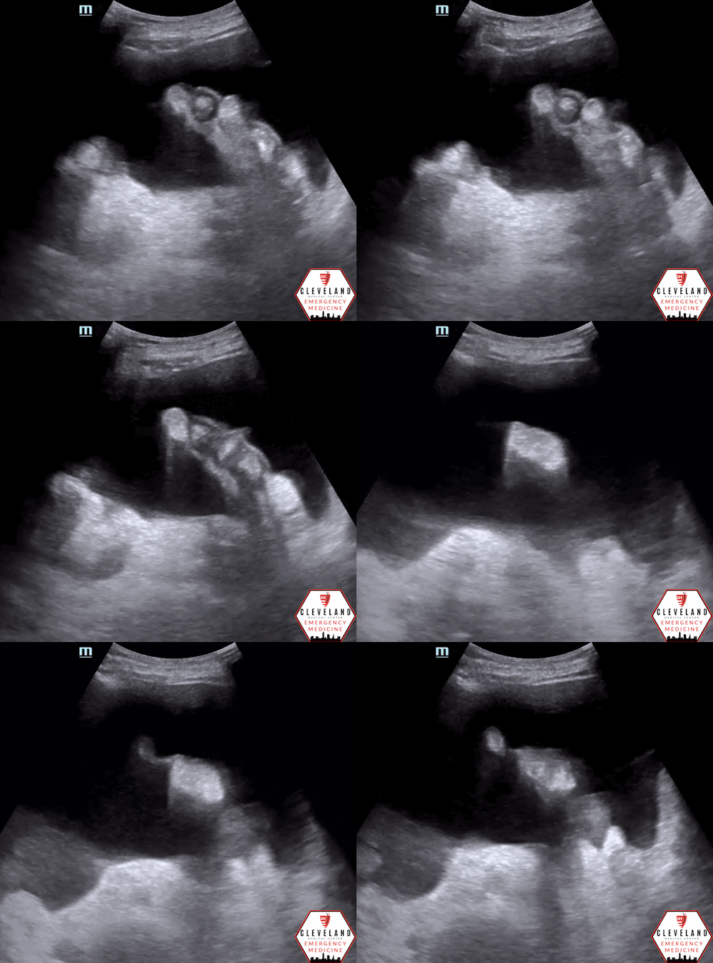
Large volume of ascites without overlying superficial vessels.
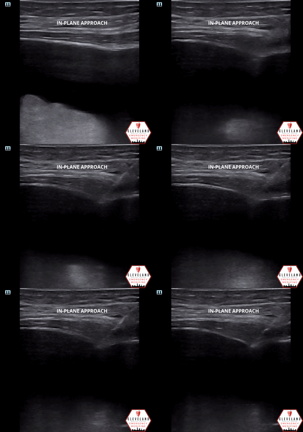
Successful paracentesis with needle visualized within the peritoneal fluid
Case Continued
The paracentesis was uncomplicated and approximately 40 cc’s of turbid yellow fluid was aspirated from the peritoneal cavity. Fluid studies, including gram stain, culture, cell count, protein, glucose, LDH, amylase, and triglyceride levels, were obtained. The patient was empirically started on vancomycin and piperacillin/tazobactam based on the appearance of the peritoneal fluid.
The fluid results demonstrated 2+ granulocytes but no organisms on gram stain; 215 WBC, 5000 RBC, 40% neutrophils, 26% lymphocytes, and 35% monocytes on cell count; a protein level of 1.1, triglyceride level of 500, amylase level of 25, glucose level of 125, and LDH level of 40. The culture eventually resulted as no growth.
Based on these results, the patient was diagnosed with chylous ascites, and antibiotics were discontinued. During his hospital stay, he underwent a therapeutic paracentesis (again with chylous ascites) and had improvement of his orthostasis after volume expansion with albumin and increasing doses of Midodrine. Additional workup, including stress echo, was unremarkable, and he was discharged a little over a week later.
POCUS in the Evaluation and Management of Ascites
Classically, providers had to rely on physical exam maneuvers for diagnosis of ascites, and they would perform paracenteses based on landmarks and physical exam. With the advent of higher quality, less expensive, and more portable ultrasound machines, point of care ultrasound has largely supplanted many of these outdated techniques. Multiple studies from the 1980s and 1990s have looked at the sensitivity, specificity, positive- and negative-likelihood ratios for various classic physical exam maneuvers such as bulging flanks, flank dullness, shifting dullness, and the presence of a fluid wave. Even in these studies, ultrasound is used as the gold standard with which each of these techniques is compared. None of the techniques have a sensitivity above 90% and fluid wave is the only one with a specificity at 90% or above (Table 1) [1-2].
Table 1. Performance of classically taught physical exam findings for the diagnosis of ascites. Adapted from Williams & Simel 1992 [1]
Early studies cite a 1970 article by Goldberg et al. stating that as little as 100 cc of peritoneal free fluid could be identified with transabdominal ultrasound. However, this study was done with cadaveric models placed in various positions and it was found that the right lateral decubitus and hands and knees positions were the most sensitive for ascities [3]. More recent studies of the sensitivity of the FAST exam suggest that the minimum amount of fluid detectable is closer to 200cc, if not more in inexperienced sonographers [4].
Evaluation of ascites
The evaluation of ascites begins with the simple yes-or-no question of whether there is intraperitoneal free fluid or not. This is done in a similar fashion to that of the abdominal views of a FAST exam. Starting from the right upper quadrant, evaluate Morrison’s pouch, the caudal tip of the liver, and the right paracolic gutter. This view is most sensitive for detecting free fluid [5]. Progressing to the other views, evaluate the splenodiaphragmatic recess and splenorenal recess in the LUQ and the rectovesicular space or rectouterine space in the pelvis [4].
Identifying Optimal Puncture Site / Minimizing Complications
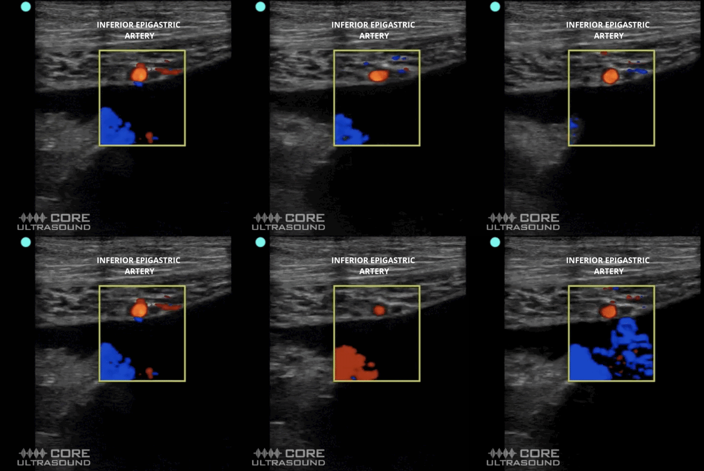
Once ascites is identified, providers should scan in a lawn-mower pattern to identify the pocket with the largest amount of free fluid. If there are indications to perform a diagnostic or therapeutic paracentesis, this is the optimal location to do so. Use color doppler to ensure there are no abdominal wall vessels, particularly the inferior epigastric vessels, at risk of injury during the procedure. Bleeding is among the most common complications.
Also make note of the abdominal wall thickness (distance from the skin surface to the peritoneal cavity) as well as the depth of the fluid pocket (or distance to bowel/organs). Using these measurements, select a needle for the procedure that is long enough to ensure access to the peritoneal cavity, but short enough to avoid damaging bowel or other intraabdominal/pelvic organs. The optimal site for puncture is where the abdominal wall is the thinnest, there’s no vasculature near the trajectory of the needle, and there’s adequate depth of fluid without intraabdominal organs nearby [6].
Source: coreultrasound.com
Color doppler to ensure no vasculature in the abdominal wall
Measure abd wall thickness & depth to bowel/organs
Ultrasound Guidance for Paracentesis
The procedure itself can be done either under static or dynamic ultrasound guidance. Static guidance utilizes the above technique of identifying the largest pocket of fluid, marking its position, and then doing the procedure “blindly.” Dynamic guidance utilizes techniques similar to that of vascular access using either an in-plane or out-of-plane approach to directly visualize and guide the needle tip into the peritoneal cavity. This technique is preferred if the fluid pocket is small, if there is are neighboring structures, such as bowel, that risk injury, and/or if the patient moves prior to or during the procedure. There are no studies demonstrating superiority of one technique over another. However, compared to the traditional landmark-based approach, ultrasound-guidance has demonstrated higher success rates, reduced complications, and lower costs, and societies have recommended the use of ultrasound for paracentesis as a result [7-10].
A Few Pearls & Pitfalls
Optimally position the patient — elevate head of bed to help draw fluid toward the lower quadrants, away from liver and spleen. Can also tilt the patient toward the side of puncture to further help move fluid to that area.
While the curvilinear probe is best for the ascites assessment, switching to the linear probe can result in better needle visualization for the actual procedure
Minimize patient (+/- probe) movement after identifying optimal site to avoid shifting of structures and puncturing bowel or other organs.
Take Home Points
POCUS is more accurate for diagnosis of all volumes of ascites than physical exam maneuvers.
Use POCUS to identify the largest fluid pocket and optimal site for paracentesis
Apply Color Doppler over the abdominal wall to ensure no vessels overlying the target fluid
Estimate abdominal wall thickness and depth of fluid to help determine appropriate needle length and reduce risk of bowel/organ injury
Ultrasound-guided paracentesis has higher success rates and fewer complications compared to the landmark-based approach. Consider dynamic guidance, especially for smaller fluid pockets
POST BY: DR. SAM HERTZ (R1)
FACULTY EDITING BY: DR. LAUREN MCCAFFERTY
References
Williams JW, Simel DL. Does This Patient Have Ascites?: How to Divine Fluid in the Abdomen. JAMA J Am Med Assoc. 1992;267(19):2645-2648. doi:10.1001/jama.1992.03480190087038
Cummings S, Papadakis M, Melnick J, Gooding GA, Tierney LM. The predictive value of physical examinations for ascites. West J Med. 1985;142(5):633-636.
Goldberg BB, Goodman GA, Clearfield HR. Evaluation of Ascites by Ultrasound. 1970;96(1):15-22. doi:10.1148/96.1.15 https://doi.org/101148/96115
Bloom BA, Gibbons RC. Focused Assessment with Sonography for Trauma. In: StatPearls. Treasure Island, FL: StatPearls Publishing. Accessed June 22, 2022. https://www.ncbi.nlm.nih.gov/books/NBK470479/
Lobo V, Hunter-Behrend M, Cullnan E, Higbee R, Phillips C, Williams S, Perera P, Gharahbaghian L. Caudal edge of the liver in the right upper quadrant (RUQ) view is the most sensitive area for free fluid on the FAST exam. West J Emerg Med. 2017; 18(2), 270–280. https://doi.org/10.5811/westjem.2016.11.30435
Ennis J, Schultz G, Perera P, Williams S, Gharahbaghian L, Mandavia D. Ultrasound for Detection of Ascites and for Guidance of the Paracentesis Procedure: Technique and Review of the Literature. International J Clin Med. 2014; 05(20): 1277–1293. https://doi.org/10.4236/ijcm.2014.520163
Ultrasound-Guided Paracentesis - ACEP Now. Accessed June 22, 2022. https://www.acepnow.com/article/ultrasound-guided-paracentesis/?singlepage=1&theme=print-friendly
Cho J, Jensen TP, Reierson K, et al. Recommendations on the Use of Ultrasound Guidance for Adult Abdominal Paracentesis: A Position Statement of the Society of Hospital Medicine. J Hosp Med. 2019;14:E7. doi:10.12788/JHM.3095
Nazeer, Shameem R., et al. “Ultrasound-Assisted Paracentesis Performed by Emergency Physicians vs the Traditional Technique: a Prospective, Randomized Study.” The American J Emerg Med. 2005; 23(3):363–367. doi:10.1016/j.ajem.2004.11.001.
Patel PA, Ernst FR, Gunnarsson CL. Evaluation of Hospital Complications and Costs Associated with Using Ultrasound Guidance during Abdominal Paracentesis Procedures. J Med Economics. 2012; 15: 1-7. http://dx.doi.org/10.3111/13696998.2011.628723


