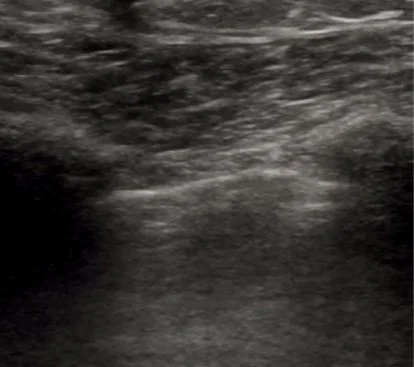
This latest Intern US of the Month is by Dr. Sam Hertz and features a great discussion on using POCUS to evaluate for ascites and guide paracentesis.
Read MoreThis latest Intern US of the Month case by Dr. Juan Valdes Infante showcases the utility of POCUS in evaluating for multiple ankle pathologies. Learn more about calcaneal avulsion fractures, which are often a surgical emergency, and how to assess for various ankle pathologies using POCUS!
Read MoreThis Intern US of the Month features a great case by Dr. Rich Dowd of an extensive aortic dissection spanning from the aortic arch to the bifurcation with extension into the left subclavian artery. Check out the full post to learn more!
Read MoreThis latest Intern US of the Month features a case of a traumatic pneumothorax and a great discussion on this time-saving and highly useful POCUS assessment by Dr. Blake Nelson!
Read MoreThis Intern US of the Month features a unique case of asteroid hyalosis, a mimicker of vitreous hemorrhage, diagnosed with POCUS by Dr. Eniola Gros!
Read MoreThis latest Intern US of the Month features a great case of small (and large) bowel obstruction and discussion on how and why to assess for this with POCUS! Brought to you by Dr. Will Heersink!
Read MoreThis is a great case and discussion by Dr. Connor Parsell (PGY2) of imperforate hymen resulting in hematocolpos, diagnosed clinically and confirmed with POCUS in the ED.
Read MoreThis month’s case by Dr. Kalee Royster (PGY1) features a great example of a pericardial effusion with developing sonographic findings of tamponade along with discussion of how to evaluate this on POCUS.
Read MoreThis month’s case by Dr. Nick Dimeo (PGY1) demonstrates how Biliary POCUS can quickly aid in clinical management/narrowing the differential/disposition and provides some great tips on how to master this exam.
Read MoreThis month’s case by Dr. Dylan Sexton features a great example of a lung mass along with a discussion of a lung ultrasound assessment for consolidation.
Read MoreThis month’s case is a complex patellar fracture diagnosed with POCUS along with a great discussion of the knee ultrasound exam by Dr. Sofia Chinchilla.
Read MoreIt’s our first Intern Ultrasound of the Month for this academic year! Here’s a great case and discussion by Dr. Bejan Kanga, PGY1, about using POCUS to diagnose shoulder dislocation and confirm reduction!
Read MoreThis Intern Ultrasound of the Month by Dr. Wes Gallaher features a great case of Achilles tendon rupture confirmed with POCUS! Read on to learn more!
Read MoreThis Intern Ultrasound of the Month is by Dr. Anna Williams. It features a great case of a DVT quickly diagnosed at the bedside, which allowed for expeditious initiation of heparin long before extensive PEs were found on CT. Also helped rapidly rule out other suspected pathology. Read on to learn more!
Read MoreThis Intern Ultrasound of the Month by is by Dr. Dan Saadeh and features a great case of a STEMI with heart block, first detected with POCUS which found regional wall motion abnormalities. EKG confirmed the diagnosis. Read on to learn more!
Read MoreThis Intern Ultrasound of the Month by Dr. Dani Rao is a great case of extensor tenosynovitis from a cat bite. Because of this POCUS diagnosis (when the clinical presentation was somewhat vague), the patient was evaluated by hand surgery and admitted for IV antibiotics. #POCUSforthewin
Read MoreThis Intern Ultrasound of the Month by Dr. Haley Wartman features a great case of IVC thrombus found incidentally when scanning the aorta and kidneys that led to new diagnosis and further workup of metastatic disease.
Read MoreThis Intern Ultrasound of the Month by Dr. Jehanne Belange features a great case of acute coronary syndrome in which POCUS detected a regional wall motion abnormality mimicking the classic Takotsubo pattern.
Read MoreThis Intern Ultrasound of the Month features a great case of retinal detachment diagnosed with POCUS by Dr. Connor Parsell, PGY1.
Read MoreThis month’s Intern Ultrasound of the Month is a great case by Dr. Mike Fellenbaum, PGY1, of large bilateral emphysematous bullae whose POCUS findings mimic those of a pneumothorax.
Read More


